Understanding Ultrasound: A Complete Guide
Oct 18, 2025Ultrasound is one of the most commonly used imaging techniques in modern medicine. It’s safe, non-invasive, and provides real-time images of organs, tissues, and blood flow inside the body. Whether you’re visiting for a routine scan or diagnostic evaluation, understanding how ultrasound works can help you appreciate the vital role it plays in medical care.
In this guide, we’ll dive deep into what ultrasound is, how it’s performed, what the results mean, and why it remains a crucial diagnostic tool across healthcare specialties.
What Is an Ultrasound?
An ultrasound, also known as sonography, is a medical imaging technique that uses high-frequency sound waves to produce images of internal structures. Unlike X-rays or CT scans, ultrasound does not use ionizing radiation, making it a safer option, particularly for pregnant women and developing babies.
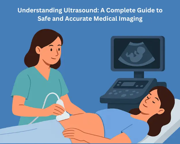
The images are generated by an instrument called a transducer, which sends sound waves into the body. These waves bounce off organs and tissues, returning echoes that the computer translates into real-time visual images on a monitor.
Step-by-Step Ultrasound Procedure Explained
Although different types of ultrasounds target different parts of the body—such as the abdomen, pelvis, heart, or blood vessels—the basic procedure follows a general pattern. Here’s what to expect:
1. Preparation
Depending on the type of ultrasound, you may be asked to:
1. Fast for a few hours, especially for abdominal scans (to reduce gas interference).
2. Drink water before pelvic scans to ensure the bladder is full for clearer images.
3. Remove jewelry or clothing from the area being examined.
2. Positioning and Application of Gel
You’ll lie down on an examination table, and the technician (called a sonographer) will apply a layer of water-based gel to your skin. The gel eliminates air pockets between the skin and the transducer, allowing sound waves to travel more efficiently.
3. Image Capture
The sonographer gently glides the transducer over the gelled area. As the transducer emits sound waves, echoes bounce back and are captured to create moving images on the screen. The process is completely painless and usually lasts 15–30 minutes depending on the area being examined.
4. During the Scan
You might be asked to change positions, hold your breath, or adjust the angle of your body to get better image quality. For specialized scans, such as transvaginal or transesophageal ultrasounds, a small probe may be inserted internally for closer imaging.
5. After the Scan
Once captured, the gel is wiped off, and you can resume normal activities immediately. There are no side effects or recovery time needed.
Decoding Ultrasound Results: What You Need to Know
After your ultrasound, the images are reviewed and interpreted by a radiologist or specialized doctor. Understanding the results can feel overwhelming, but here’s what’s typically involved:
1. Normal Findings
A normal ultrasound shows organs that are uniform in shape, size, and structure, without signs of irregular growths, blockages, fluid buildup, or abnormal motion. Blood flow appears consistent, and tissues reflect sound evenly.
2. Abnormal Findings
Depending on the part of the body examined, abnormal ultrasound findings may include:
1. Cysts or fluid collections
2. Tumors or solid masses
3. Inflammation or infection (shown by tissue swelling)
4. Abnormal blood flow (detected using Doppler ultrasound)
5. Pregnancy complications or fetal growth issues
The radiologist will compile these findings in a detailed report, which your doctor reviews to explain what the results mean for your diagnosis or treatment plan.
3. Doppler Ultrasound Insights
A Doppler ultrasound specifically measures how blood moves through your vessels and heart. It helps detect:
1. Blocked arteries or veins (such as in deep vein thrombosis)
2. Reduced blood flow to organs
3. Leaky or narrowed blood vessels
Doppler studies are essential for cardiovascular assessments, offering crucial information without the need for invasive tests.
Types of Ultrasound Scans
Ultrasound is incredibly versatile, with different types tailored for various medical needs:
1. Abdominal Ultrasound – Evaluates organs such as the liver, gallbladder, kidneys, pancreas, and spleen.
2. Pelvic Ultrasound – Commonly used for reproductive organs in both men and women; critical during pregnancy.
3. Obstetric Ultrasound – Monitors fetal development and maternal health throughout pregnancy.
4. Cardiac Ultrasound (Echocardiogram) – Examines heart chambers, valves, and pumping efficiency.
5. Vascular Ultrasound – Assesses blood flow in veins and arteries across the body.
6. Musculoskeletal Ultrasound – Evaluates muscles, tendons, and joints for tears or inflammation.
7. Each category of ultrasound provides valuable insights for accurate diagnosis and treatment planning.
Why Ultrasound Is a Vital Diagnostic Tool
Ultrasound plays an integral part in modern diagnosis because it combines safety, accuracy, and convenience.
1. Safe and Non-Invasive
Since ultrasound uses sound waves instead of radiation, it poses virtually no health risk—even for pregnant women and children. It is widely preferred for prenatal imaging because it protects both mother and baby.
2. Real-Time Imaging
Ultrasound produces live images, allowing doctors to visualize motion in organs, blood flow, or the movement of a fetus. This makes it invaluable for guiding procedures like biopsies and injections.
3. Portable and Cost-Effective
Unlike large imaging systems such as MRI or CT scanners, ultrasound machines are portable and more affordable, making them vital in both hospitals and rural clinics where quick decisions are needed.
4. Versatile Applications
From cardiac care and obstetrics to musculoskeletal and emergency medicine, ultrasound supports various specialties. Surgeons even use intraoperative ultrasound during surgeries to guide complex procedures.
5. Early Detection and Continuous Monitoring
Ultrasound allows doctors to detect problems early and track treatment progress without exposing patients to harmful radiation. Long-term monitoring for conditions like liver disease, heart failure, or cancer follow-ups can be done safely.
How to Prepare for an Ultrasound
Preparation depends on the imaging area, but some general tips include:
1. Follow fasting instructions if required.
2. Wear loose, comfortable clothing.
3. Arrive on time to fill out any necessary medical forms.
4. Bring prior imaging reports for comparison.
For pregnancy ultrasounds, patients might be advised to maintain a full bladder to improve visibility.
The Role of Ultrasound in Modern Healthcare
Ultrasound isn't just diagnostic—it’s also therapeutic and interventional. With new advancements like 3D and 4D imaging and point-of-care ultrasound (POCUS) used by emergency physicians, its role continues to expand.
Here are a few areas where ultrasound is transforming care:
1. Emergency medicine: Quick detection of internal bleeding or organ damage during trauma care.
2. Guided procedures: Used for biopsies, injections, and catheter placements.
3. Prenatal monitoring: Fetal growth, organ formation, and placental position assessments.
4. Cardiac imaging: Detecting structural defects or heart failure early on.
Key Takeaways
1. Ultrasound is safe, radiation-free, and highly versatile. It effectively diagnoses conditions involving soft tissues and vascular structures.
2. Simple preparation and real-time results make it an ideal first-line diagnostic tool.
3. Interpreting ultrasound results correctly helps in tailoring accurate treatment plans.
4. Technological advancements like Doppler and 3D imaging continue to expand its diagnostic power.
Conclusion
Ultrasound imaging remains an essential cornerstone of modern diagnostics. Whether it’s evaluating your heart, monitoring fetal health, or identifying internal conditions, ultrasound offers quick, clear, and safe insight into your body’s inner workings.
If your doctor has recommsended an ultrasound, you can feel confident knowing it’s one of the most trusted, reliable, and safe medical imaging methods available today.

